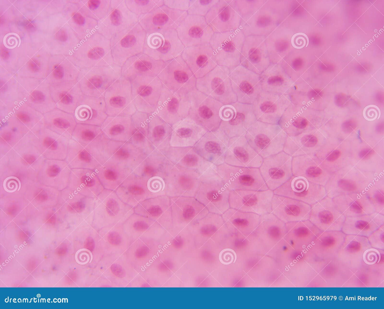
Animal Cell Visible Under A Light Microscope : Cell Structure Organisation Cie Igcse Biology Revision Notes : Living specimens can be viewed in their natural colour, although stains may be applied to resolve specific structures.
byLaurie Veith-0
Animal Cell Visible Under A Light Microscope : Cell Structure Organisation Cie Igcse Biology Revision Notes : Living specimens can be viewed in their natural colour, although stains may be applied to resolve specific structures.. Explanation:the two visible structures that are visible with 'light microscope' are the cell wall and the vacuoles. Living specimens can be viewed in their natural colour, although stains may be applied to resolve specific structures. Cells consist of cytoplasm enclosed within a membrane, which contains many biomolecules such as proteins and nucleic acids.2 most plant and animal cells are only visible under a light microscope, with dimensions between 1 and 100 micrometres.3 electron microscopy gives a. An animal and plant cell as seen under a light microscope. Some organelles are visible with a compound light microscope, while other organelles can be seen only under a more powerful tool, such as an electron microscope.
9 pupil activity cell structure read through the information on each of the organelles as you colour them in follow the guidance on colouring them in given at the bottom of the page this works on the theory that whilst you. The plant cells have cell wall that is made up of cellulose and the cell membrane is present beneath this cell wall whereas the animal cell contains only the cell membrane. The organelles in a cheek cell that are not visible under a light microscope are the ribosomes. Plant and animal cells have a nucleus inside the cytoplasm. A cell is a very tiny structure which exists in living bodies.
Course S4 Biology Topic Unit 3 Microscopy from elearning.reb.rw Mere magnification without added detail is. See how a generalized structure of an animal cell and plant cell look with labeled diagrams. Is ribosome visible under light microscope? Cell is a tiny structure and functional unit of a living organism containing various parts known as organelles. Under a light microscope, the cell membrane, nucleus and cytoplasm of a cheek cell (animal cell) can be observed. The sister chromatids begin to coil more tightly with the aid of condensin proteins and become visible under a light microscope. Electron microscopes use accelerated electron beams (as opposed to visible light in a light microscope) to create images of magnification as high as 1 million x and has a very high individual cells are easily visible under both visible and electron microscopy of sufficient magnification. 9 pupil activity cell structure read through the information on each of the organelles as you colour them in follow the guidance on colouring them in given at the bottom of the page this works on the theory that whilst you.
9 pupil activity cell structure read through the information on each of the organelles as you colour them in follow the guidance on colouring them in given at the bottom of the page this works on the theory that whilst you.
Animal cells are typical of the eukaryotic cell, enclosed by a plasma membrane and containing a the lack of a rigid cell wall allowed animals to develop a greater diversity of cell types, tissues, and the microscope has been a fundamental tool in the field of cell biology and is often used to observe. It can be used to view dead and living samples and can maximize these samples up to one thousand times their actual size. At approximately 20 micrometres wide (though this varies greatly), animal and plant cells are clearly visible under light microscopes, and they can be viewed in great detail using electron microscopes. Electron microscopes use accelerated electron beams (as opposed to visible light in a light microscope) to create images of magnification as high as 1 million x and has a very high individual cells are easily visible under both visible and electron microscopy of sufficient magnification. These include the cell membrane, cytoplasm and the nucleus. The plant cell as more rigid and stiff walls. The sister chromatids begin to coil more tightly with the aid of condensin proteins and become visible under a light microscope. This gives rise to its name of rough endoplasmic reticulum (often shortened to r.e.r.) Below the basic structure is shown in the same animal cell, on the left viewed with the light microscope, and on the right with the transmission electron microscope. Nerve cells under light microscope nerve cell of spinal cord, onion epidermis with large cells under light microscope, ppt eukaryotic cell seen. The organelles in a cheek cell that are not visible under a light microscope are the ribosomes. Some features common to animal cells. Magnification, however, is not the most important issue in microscopy.
Mere magnification without added detail is. In most plant cells, the organelles that are visible under a compound {light} microscope are the cell wall, cell membrane, cytoplasm the animal cell is more fluid or elastic or malleable in structure; People's surprise, mitochondria are visible in the light microscope. The light microscope employs visible light, to detect small objects and is the well known and well used research tool in biology. The plant cells have cell wall that is made up of cellulose and the cell membrane is present beneath this cell wall whereas the animal cell contains only the cell membrane.
17 385 Animal Cell Photos Free Royalty Free Stock Photos From Dreamstime from thumbs.dreamstime.com Light microscopes using visible light and lenses to form a magnified image of the object under investigation e.g. Cell structure teaching resources the science teacher. In most plant cells, the organelles that are visible under a compound {light} microscope are the cell wall, cell membrane, cytoplasm the animal cell is more fluid or elastic or malleable in structure; A compound light microscopes use lenses and light to magnify cell parts. 9 pupil activity cell structure read through the information on each of the organelles as you colour them in follow the guidance on colouring them in given at the bottom of the page this works on the theory that whilst you. Electron microscopes use accelerated electron beams (as opposed to visible light in a light microscope) to create images of magnification as high as 1 million x and has a very high individual cells are easily visible under both visible and electron microscopy of sufficient magnification. Explain that cells take in nutrients in order to grow, divide and to make needed materials. However, they usually can achieve a how can you tell the difference between plant and animal cells under a microscope?
See how a generalized structure of an animal cell and plant cell look with labeled diagrams.
Is ribosome visible under light microscope? Most cells are visible under a light microscope, but mitochondria and bacteria are barely visible. It controls all the processes and chemical reactions that. It can be used to view dead and living samples and can maximize these samples up to one thousand times their actual size. With a light microscope you can see individual cells and large subcellular structures like the nucleus, but not internal cell structures such as ribosomes or. 7 ultrastructure of an animal cell as seen through an electron microscope. Living specimens can be viewed in their natural colour, although stains may be applied to resolve specific structures. The organelles in a cheek cell that are not visible under a light microscope are the ribosomes. Animal and plant cell lab. Some organelles are visible with a compound light microscope, while other organelles can be seen only under a more powerful tool, such as an electron microscope. Faintly visible are several mitochondria, for example the grey oval structures at the bottom left. You can see yeast cells, animal cells, and plant cells pretty well with a 400x magnification you will need an electron microscope to see the viruses. Electron microscopes use accelerated electron beams (as opposed to visible light in a light microscope) to create images of magnification as high as 1 million x and has a very high individual cells are easily visible under both visible and electron microscopy of sufficient magnification.
Nerve cells under light microscope nerve cell of spinal cord, onion epidermis with large cells under light microscope, ppt eukaryotic cell seen. Plant and animal cells a. Cells consist of cytoplasm enclosed within a membrane, which contains many biomolecules such as proteins and nucleic acids.2 most plant and animal cells are only visible under a light microscope, with dimensions between 1 and 100 micrometres.3 electron microscopy gives a. As per the given information in the question, cells of mushrooms, plants, and animals all have visible nuclei under a microscope. However, they usually can achieve a how can you tell the difference between plant and animal cells under a microscope?
Basic Parts Of A Cell And Cell Organelles Ck 12 Foundation from dr282zn36sxxg.cloudfront.net These organelles are responsible for protein synthesis. An animal and plant cell as seen under a light microscope. You can see yeast cells, animal cells, and plant cells pretty well with a 400x magnification you will need an electron microscope to see the viruses. See how a generalized structure of an animal cell and plant cell look with labeled diagrams. The original electron microscopic image of viruses. These are both specific types of cells, and from specific species. Cells consist of cytoplasm enclosed within a membrane, which contains many biomolecules such as proteins and nucleic acids.2 most plant and animal cells are only visible under a light microscope, with dimensions between 1 and 100 micrometres.3 electron microscopy gives a. It controls all the processes and chemical reactions that.
Endoplasmic reticulum studded with ribosomes looks rough under the microscope;
Is ribosome visible under light microscope? In most plant cells, the organelles that are visible under a compound {light} microscope are the cell wall, cell membrane, cytoplasm the animal cell is more fluid or elastic or malleable in structure; Observing a wide range of biological processes and animal cell under light microscope is easier due to advances in microscopic techniques. Cell structure teaching resources the science teacher. Types of light microscope 1. However, they usually can achieve a maximum of 2000x magnification which is not sufficient to see many other tiny organelles like ribosomes, endoplasmic reticulum, lysosomes, centrioles, golgi bodies unless they have an electron. The plant cell as more rigid and stiff walls. Some organelles are visible with a compound light microscope, while other organelles can be seen only under a more powerful tool, such as an electron microscope. Explanation:the two visible structures that are visible with 'light microscope' are the cell wall and the vacuoles. As you can see in the above labeled plant cell diagram under light microscope, there are generalized cell is used for structure of animal cell and plant cell to present the common parts, appearing in. Cells consist of cytoplasm enclosed within a membrane, which contains many biomolecules such as proteins and nucleic acids.2 most plant and animal cells are only visible under a light microscope, with dimensions between 1 and 100 micrometres.3 electron microscopy gives a. Some features common to animal cells. Mere magnification without added detail is.


Post a Comment